Lower Body Bone Diagram / Bones Of The Lower Limb Anatomy And Physiology I / The lumbar spine makes up the the lower end of the spinal column.. This diagram depicts lower extremity diagram.human anatomy diagrams show internal organs, cells, systems, conditions, symptoms and sickness information and/or tips for healthy living. Bone diagram forehead (frontal bone) nose bones (nasals) cheek bone (zygoma) upper jaw (maxilla) lower jaw (mandible) breast bone (sternum) upper arm bone (humerus) lower arm bone (ulna) thigh bone (femur) collar bone (clavicle) toe bones (phalanges) ankle bones. The lower left quadrant of the abdomen is complex leaving its many structures prone to inflammation obstruction or injury. This diagram depicts lower extremity muscles diagram.human anatomy diagrams show internal organs, cells, systems, conditions, symptoms and sickness information and/or tips for healthy living. The body is cylindrical in its upper portion, and more prismatic below.
The long bones of the body contain many distinct regions due to the way in which they develop. The lumbar and sacrum region make up the bone of the lower back anatomy. The vertebral column of the lower back includes the five lumbar vertebrae, the sacrum, and the coccyx. They hold up your body, and along with your muscles, keep you moving. Other sesamoid bones can form in the joints of the hands and feet, but are not present in all people.
1 from It has an upper extremity, a shaft, and a lower extremity, all of which are full of various structural landmarks. For more anatomy content please follow us and visit our website: Your lower back (lumbar spine) is the anatomic region between your lowest rib and the upper part of the buttock. This article looks at the anatomy of the back, including bones, muscles. Also called the shin bone, the tibia is the longer of the two bones in the. The bones of the pelvis and lower back work together to support the body's weight, anchor the abdominal and hip muscles, and protect the delicate vital organs of the vertebral and abdominopelvic cavities. The l5 vertebra is connected to the top of. L1, l2, l3, l4, and l5.
Your lower back (lumbar spine) is the anatomic region between your lowest rib and the upper part of the buttock.
The anatomy of the lumbar spine is quite complex. Other sesamoid bones can form in the joints of the hands and feet, but are not present in all people. Other pelvic muscles, such as the psoas major and iliacus, serve as flexors of the trunk and thigh at the hip joint and. The tibia (shin bone) is the medial bone of the leg and is larger than the fibula, with which it is paired (figure 6.52). They hold up your body, and along with your muscles, keep you moving. The lower leg contains two major long bones, the tibia and the fibula, which are both very strong skeletal structures. Also called the shin bone, the tibia is the longer of the two bones in the. The vertebral column of the lower back includes the five lumbar vertebrae, the sacrum, and the coccyx. Bone diagram forehead (frontal bone) nose bones (nasals) cheek bone (zygoma) upper jaw (maxilla) lower jaw (mandible) breast bone (sternum) upper arm bone (humerus) lower arm bone (ulna) thigh bone (femur) collar bone (clavicle) toe bones (phalanges) ankle bones. This is the longest bone in the human body, and is also known as the thigh bone. Related posts of anatomy of left abdomen human body bones diagram. The knee joint is the largest joint in the body and is primarily a hinge joint, although some sliding and rotation occur. The lower left quadrant of the abdomen is complex leaving its many structures prone to inflammation obstruction or injury.
The arch surrounds the hollow vertebral foramen and connects the body to the bony processes on the posterior of the vertebra. Muscle charts of the human body for your reference value these charts show the major superficial and deep muscles of the human body. The long bones of the body contain many distinct regions due to the way in which they develop. This area is commonly referred to as the calf. This curve, called lordosis, helps to:
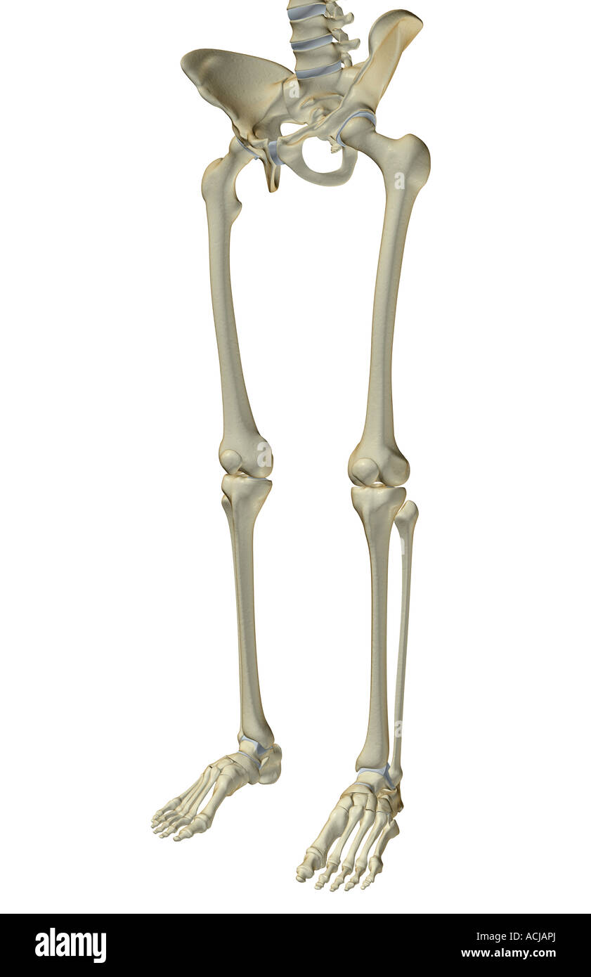
The Bones Of The Lower Body Stock Photo Alamy from c8.alamy.com Other pelvic muscles, such as the psoas major and iliacus, serve as flexors of the trunk and thigh at the hip joint and. It consists of 5 lumbar vertebra that are numbered 1 through 5 from top to bottom i.e. This is the longest bone in the human body, and is also known as the thigh bone. The back supports the weight of the body, allowing for flexible movement while protecting vital organs and nerve structures. Balance the weight of your head on top of your spine. Muscle charts of the human body for your reference value these charts show the major superficial and deep muscles of the human body. The lower extremity consists of 2 epicondyles, 2 processes (trochlea & capitulum), and 3 fossae (radial fossa, coronoid fossa, and olecranon fossa). Evenly distribute weights from your upper body into the lower extremities.
The bones of the legs are those that make up the thigh, the lower half of the legs, and the feet.
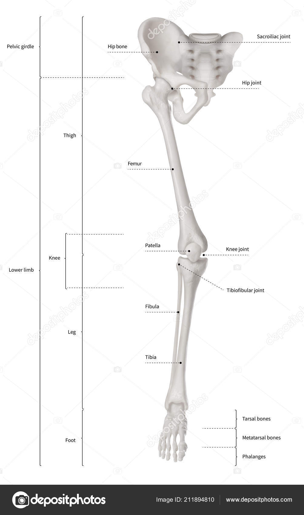
This article looks at the anatomy of the back, including bones, muscles. 1 your spine in this region has a natural inward curve. Understanding lower back anatomy is key to understanding the root of lower back and hip pain. The arch surrounds the hollow vertebral foramen and connects the body to the bony processes on the posterior of the vertebra. The bones of the legs are those that make up the thigh, the lower half of the legs, and the feet. The back supports the weight of the body, allowing for flexible movement while protecting vital organs and nerve structures. Teachme anatomy part of the teachme series the medical information on this site is provided as an information resource only, and is not to be used or relied on for any diagnostic or treatment purposes. The bones of the leg are the femur, tibia, fibula and patella.the foot bones shown in this diagram are the talus, navicular, cuneiform, cuboid, metatarsals and calcaneus. Posteriorly the body is connected to a thin ring of bone known as the arch. The vertebral column of the lower back includes the five lumbar vertebrae, the sacrum, and the coccyx. The anatomy of the lumbar spine is quite complex. The myology of the lower limb is also particularly well represented in this atlas of anatomy, with multiple anatomical charts and diagrams: The human spine is composed of 4 sections of vertebrae.
The anatomy of the lumbar spine is quite complex. 1 your spine in this region has a natural inward curve. The lower extremity consists of 2 epicondyles, 2 processes (trochlea & capitulum), and 3 fossae (radial fossa, coronoid fossa, and olecranon fossa). They hold up your body, and along with your muscles, keep you moving. The long bones of the body contain many distinct regions due to the way in which they develop.
Infographic Diagram Human Skeleton Lower Limb Anatomy Bone System Leg Stock Photo By C K Intarapong Gmail Com 211894810 from st4.depositphotos.com The patella and the pisiform bone of the carpals are the only sesamoid bones that are counted as part of the 206 bones of the body. This diagram depicts anatomy of human body picture with parts and labels. Posted on june 12, 2016 by admin. L1, l2, l3, l4, and l5. The bones of the leg are the femur, tibia, fibula and patella.the foot bones shown in this diagram are the talus, navicular, cuneiform, cuboid, metatarsals and calcaneus. Other pelvic muscles, such as the psoas major and iliacus, serve as flexors of the trunk and thigh at the hip joint and. Muscle charts of the human body for your reference value these charts show the major superficial and deep muscles of the human body. Evenly distribute weights from your upper body into the lower extremities.
Because of the important organs situated in the abdominal area, many health concerns stem.
The anatomy of the lumbar spine is quite complex. Understanding lower back anatomy is key to understanding the root of lower back and hip pain. These sections are cervical (neck), thoracic (upper and middle back), lumbar (lower back), and sacrum (tailbone). Related posts of anatomy of left abdomen human body bones diagram. These muscles, including the gluteus maximus and the hamstrings, extend the thigh at the hip in support of the body's weight and propulsion. Key bones in the abdominal area include the base of the ribcage and the lumbar spine in the lower back. For more anatomy content please follow us and visit our website: This diagram depicts lower extremity diagram.human anatomy diagrams show internal organs, cells, systems, conditions, symptoms and sickness information and/or tips for healthy living. Leg muscles anatomy human muscle anatomy muscular system anatomy anatomy bones human anatomy and physiology body anatomy leg muscles diagram muscle diagram lower leg muscles. The muscles of the lower back help stabilize, rotate, flex, and extend the spinal column, which is a bony tower of 24 vertebrae that gives the body structure and houses the spinal cord. The scaffold of the thigh is provided by the femur, the only bone of this region and the longest bone in the body. Using a median sagittal and a transverse plane passing the umbilicus at. The lower left quadrant of the abdomen is complex leaving its many structures prone to inflammation obstruction or injury.
Using a median sagittal and a transverse plane passing the umbilicus at lower body diagram. Using a median sagittal and a transverse plane passing the umbilicus at.


0 Komentar